Determination sheet1 September 2026Copenhagen, Denmark — indoors44 photographs
A crane fly on a wall, keyed to Tipula paludosa
A single male crane fly, photographed on an interior wall over about six hours with a
phone. This is the determination in full: what each character rules in or out, what the photographs can bound about its age, and where the evidence stops.
FamilyTipulidaeCertain
GenusTipulaCertain
SexMaleCertain
SpeciesT. paludosaProbable
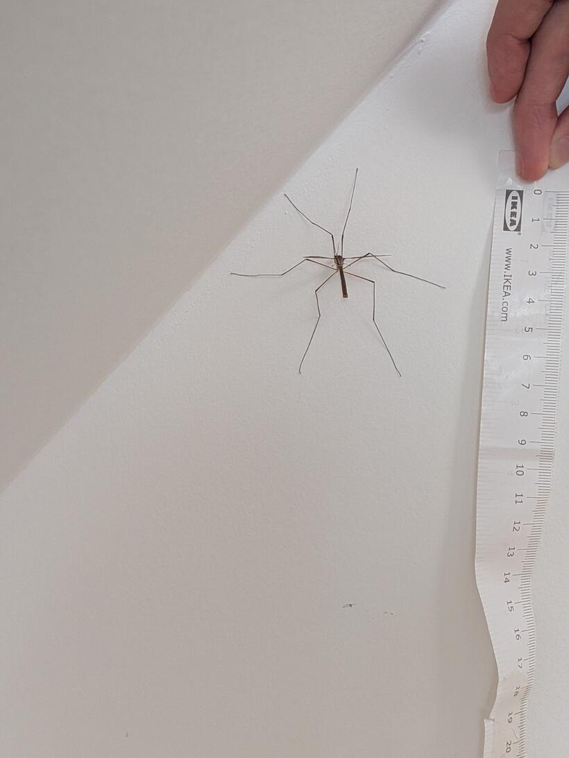
Body length≈ 20 mmmeasured against a ruler
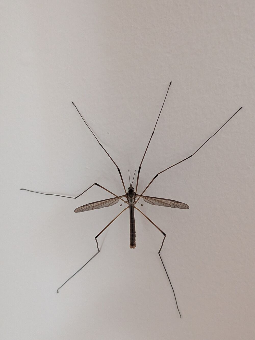
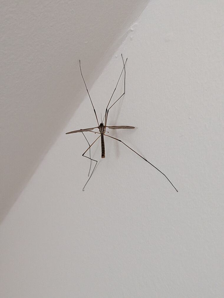
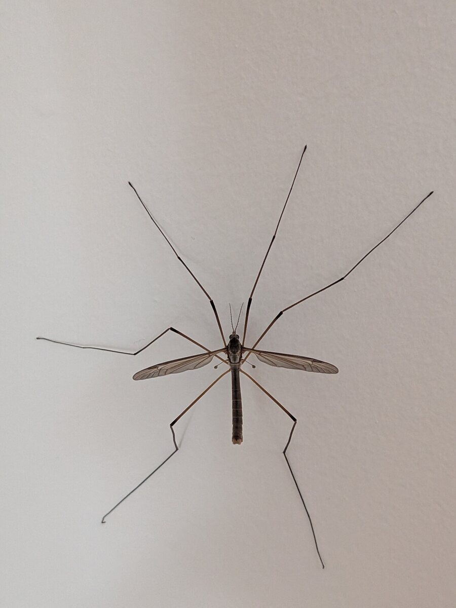
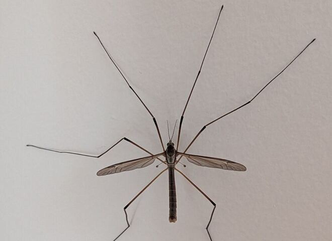
The specimen. Dorsal view, 10:24. Grey-brown, plain smoky wings held spread at rest, a darker stripe along the abdomen.First frame. 09:41, near the ceiling. Enough to place the family; nothing below it.
Running the key
order to genus
A key is a sequence of forced choices, so these steps are numbered: each only makes sense
once the one above it is settled. Green dots mark characters visible in the photographs;
red squares mark characters that rule something out.
01
Order
Diptera — the true flies
Two features settle the order together, and both are visible in a single macro frame:
One pair of wings. Everything else radiating from the thorax is a leg.
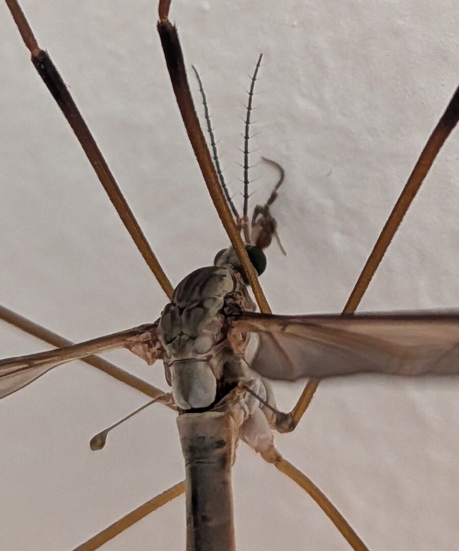
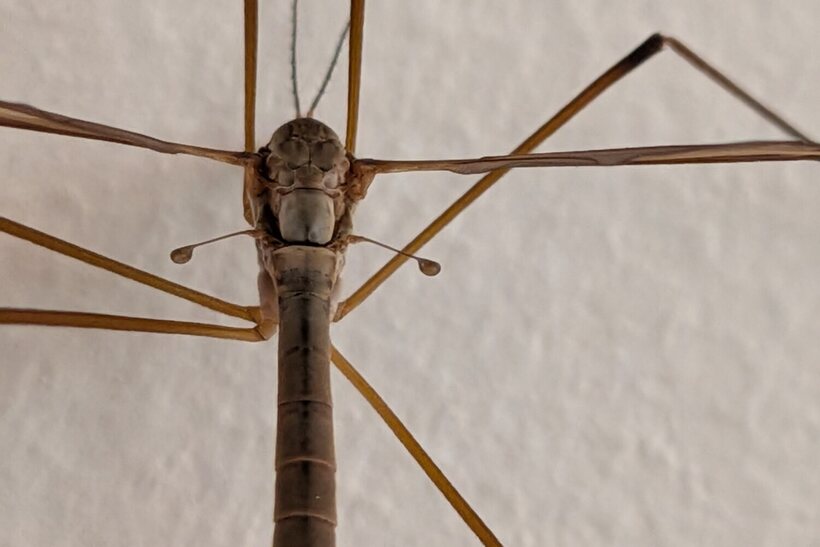
Halteres present. The small drumstick-shaped knobs behind the wing bases are the reduced hindwings — the defining dipteran character.
Plate 1. Head and thorax, 12:20. The pale drumstick at left is a haltere. Note also the bottle-green eye and the short snout (nasus).
02
Family
Tipulidae — crane flies
Long fragile legs are the obvious cue; the wing and head are what make it safe. The
characters below follow the family diagnosis in the
Cranefly Recording Scheme's introduction.
Very long, easily-shed legs with visible tibia–tarsus breaks.
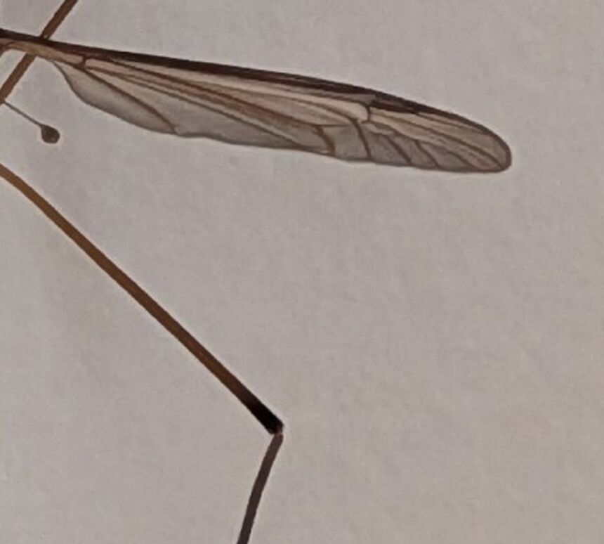
A dark pterostigma — the elongate brown patch on the leading edge near the wing tip.
Long narrow wings held spread at rest, with the discal cell and branched venation typical of the family.
A nasus — the small snout-like projection on the front of the head, visible in Plate 1.
Plate 2. Right wing. Smoky membrane, pale streak along the costa, dark pterostigma near the tip.
03
Genus
Tipula — and a large one
Scale does real work here. Measured against a ruler in the same plane, at roughly
7.3 pixels per millimetre, the body runs to about 20 mm head to abdomen tip,
with a leg span several times that.
That places it in Tipula sensu stricto — the large, grey-brown, house-entering
crane flies — rather than Nephrotoma, which would be boldly yellow and black,
or any of the small limoniids.
Plate 3. Scale frame, 10:33. The ruler sits in a slightly different plane, so treat 20 mm as ±2 — enough for genus, not for anything finer.
Sexing the specimen
a prerequisite, not a detail
Sex has to be established before the species characters mean anything, because several of
the standard couplets in Tipula apply to one sex only. Here the abdomen tip answers
it immediately.
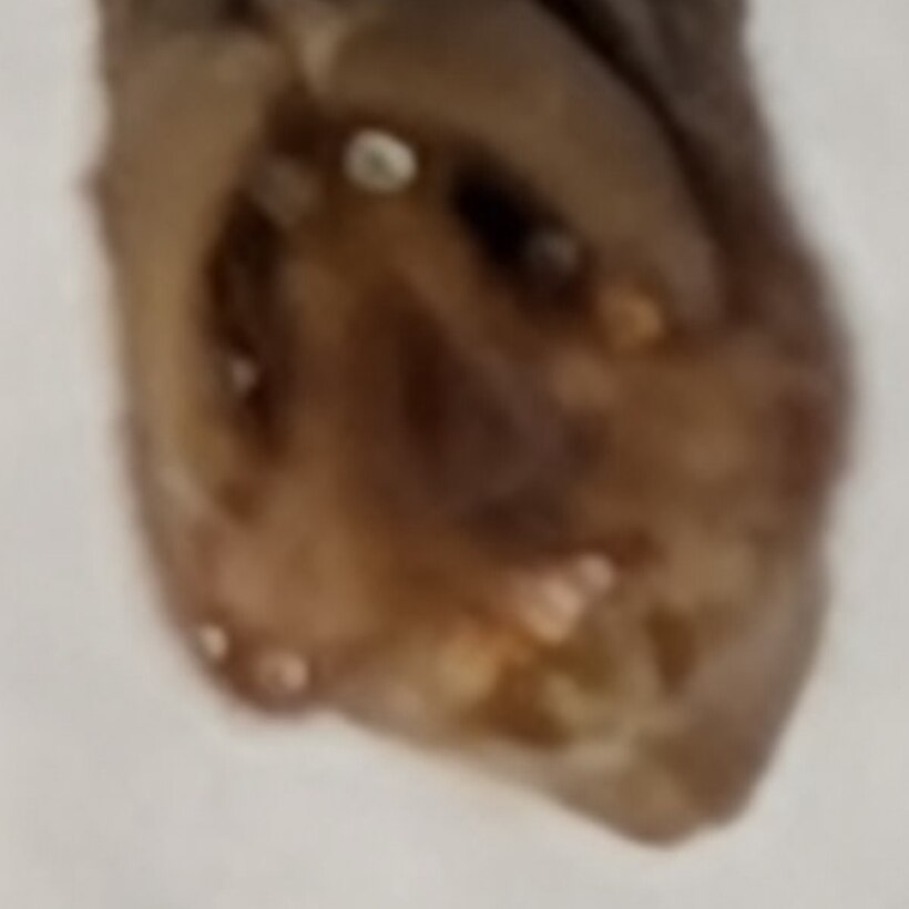
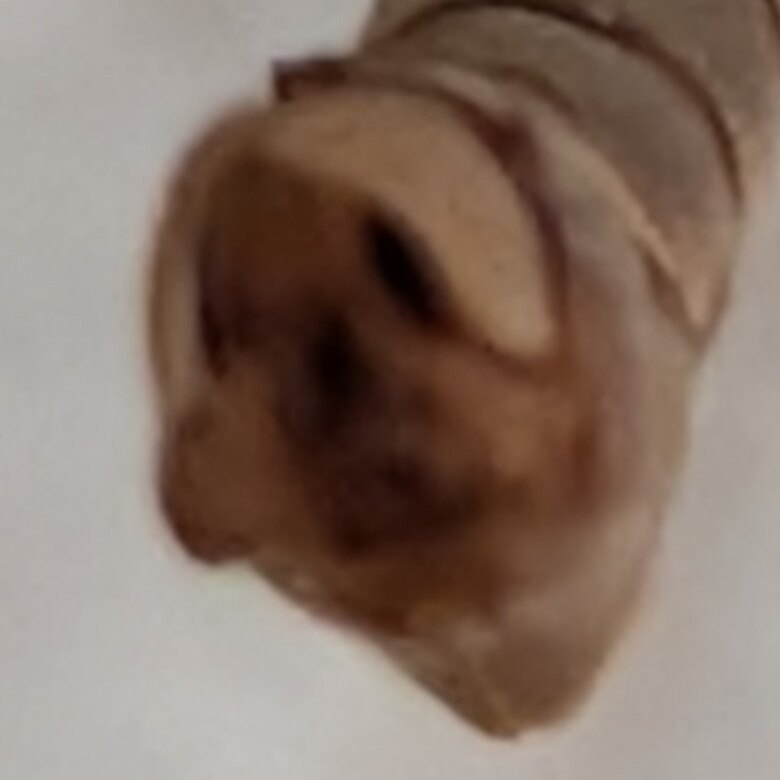
Plate 4a. Hypopygium, 15:31, close to face-on: a dark central cavity ringed by pale points at the rim. The hard white oval at the top is a flash highlight on glossy cuticle, not a structure.Plate 4b. The same capsule more obliquely, better for its profile: a pale dorsal lobe, a dark diagonal cleft, a rounded notched lower lobe.
The abdomen ends in a blunt, swollen, glossy chestnut capsule — a hypopygium, the
male genital capsule. A female would taper to a hardened, upcurved ovipositor, which is
conspicuously absent. This is the single most common sticking point when identifying crane
flies from photographs: the abdomen tip is the most obvious thing about them and it is
strongly sexually dimorphic, so a male compared against a photograph of a female reads as
damaged rather than as the other sex.
Why this doesn't reach a species
Keys do not ask whether the hypopygium is swollen and chestnut. They ask about the shapes
of the paired claspers — the gonostyli — and the sclerite between them, as set out
in Stubbs & Kramer's key to
Tipula. Plate 4a reaches the right aspect but not the resolution: once one
bright point on the rim is known to be a reflection, none of the others can be assigned
with confidence.
The antennal count
the character that decides it
With the specimen established as a large male Tipula, one countable character
separates Tipula paludosa from the rest of the genus.
13 antennal segments is the norm across the genus Tipula; paludosa,
with 14, is the exception (NatureSpot;
genus account).
It is worth being exact about what that means: the count is a test for paludosa against
the whole genus, not a two-way choice between paludosa and any single other
species. Treating it as the latter is an easy and consequential mistake.
The wing-length character sometimes quoted alongside it — wings shorter than the abdomen in
paludosa, as long as the abdomen in T. oleracea — separates females
and is no help for a male.
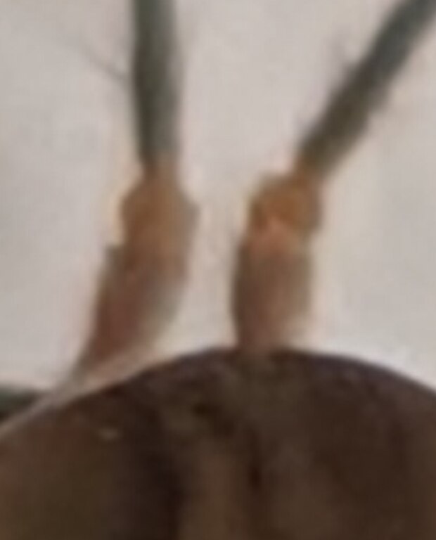
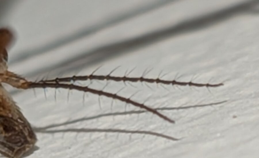
Plate 5a. Antenna bases, 15:59. Scape and pedicel are both pale tan; the flagellum begins abruptly and is dark. The pale/dark boundary is therefore the pedicel–flagellum junction.Plate 5b. Both antennae, rotated so the near one lies horizontal. They share an origin and diverge — the far one descends to the right. Only the near antenna is counted.
Plate 5c. The near antenna alone, straightened. Blue: start of the dark flagellum, and the tip. Eleven red ticks between them, each on a verticil-bearing joint. The far antenna is visible below and carries no tick.
Eleven joints delimit twelve flagellomeres. The first is plainly the longest — about
1.5× its neighbour — which is the detail that makes the base interpretable at all: an
unusually short dark piece at the base of an antenna is a pedicel caught in shadow,
not a first flagellomere.
Twelve flagellomeres + scape + pedicel = 14
Fourteen antennal segments is the character that marks out Tipula paludosa. This is
a measured count on a frame where the base, the joints and the tip are all resolved, and
on the antenna nearest the camera with the second one excluded from the field.
This count is unusually sensitive to which frame it is taken from, in a way worth stating
plainly. A backlit antenna darkens the pale pedicel until it reads as part of the flagellum,
adding a segment that is not there. An antenna angled away from the camera foreshortens the
basal segments, compressing exactly the region where the boundary has to be judged. Both
errors push the count the same way. The frame used here avoids both: the antenna lies across
the plane of the wall, side-lit, sharp from base to tip.
Character
T. paludosa
Rest of genus
This specimen
Antennal segments
14
13
12 flagellomeres → 14
Female wing length
shorter than abdomen
varies
n/a — male
Peak flight
late Aug – Sept, one generation
varies
1 September
Abundance indoors, September
the annual mass emergence
scattered
—
The date corroborates rather than carries the determination. The first of September sits on
the peak emergence of T. paludosa, which has one generation a year and produces the
familiar wave of large crane flies entering houses across northern Europe at exactly this
time (Cornell CALS).
That is an argument from base rates; on its own it would not be enough, and it is precisely
the reasoning behind a great many misidentifications — plain-winged beige crane flies are
routinely filed under paludosa by default
(iNaturalist taxon flag).
Held at probable rather than confirmed for one reason: the terminal flagellomeres
shorten toward the tip, and an error of ±1 there cannot be excluded from a photograph. A
specimen under a hand lens would remove it.
Age
bounded, not measured
No character in a photograph gives an insect's age in days. What the frames do support is a
bracket, read from wear — and for this species the bracket is narrow, because the adult
stage is very short.
Tipula paludosa adults do not feed. They emerge on the reserves laid down by the
larva, and live roughly 7–14 days; mating and most egg-laying happen within the first
24 hours (Cornell CALS;
Northeastern IPM Center).
Nearly the whole life of the animal is spent as a larva in soil; the adult is a brief
dispersal-and-mating stage.
Not newly emerged. Two things put a floor under the estimate:
The cuticle is fully hardened and pigmented. The thoracic pattern is defined and the abdominal banding crisp. A teneral crane fly is pale, soft and glossy for some hours after emergence.
The wings are fully expanded and set, with the venation hardened and the pterostigma developed.
Not old either. The wear proxies are all at zero:
All six legs present, with intact tarsi. This is the strongest of the four. Crane flies autotomise legs at the slightest provocation, and losses accumulate and are never repaired — a complete set implies a short and undisturbed adult life.
Both wings entire. Margins smooth, no nicks, tears or fraying; the membrane is clean rather than greased or darkened.
Both halteres present — small, exposed and easily lost.
Antennal verticils complete, with no broken or missing bristles along either flagellum.
Estimate
An early-adult male, most likely one to four days past emergence and within the
first half of its adult life. The date supports this independently: the first of September
sits at the beginning of the species' emergence period rather than at the end of it.
Two limits on that. Wear is a proxy, not a clock — it accumulates by chance as much as by
time, so an unlucky fresh adult can lose a leg on its first night and a sheltered one can
stay pristine longer than average. And this individual was indoors, on a smooth wall, out of
wind and rain, which is a low-wear environment; the proxies will therefore tend to read
younger than they would for an animal of the same age outdoors. The bracket should be taken
as the direction of the evidence, not a measurement.
Plate 6a. Condition, dorsal. All six legs present with intact tarsi; both wings entire and unfrayed. Compare with worn individuals, which typically show missing legs first.Plate 6b. Condition, thorax. Fully hardened and pigmented cuticle with a defined pattern, and both halteres intact.
Against reference specimens
identified material, both candidate species
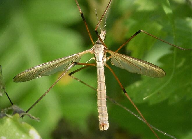
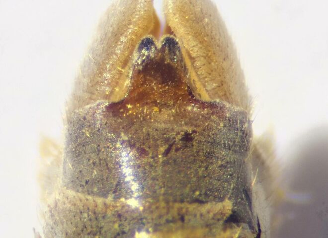
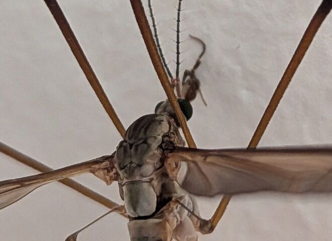
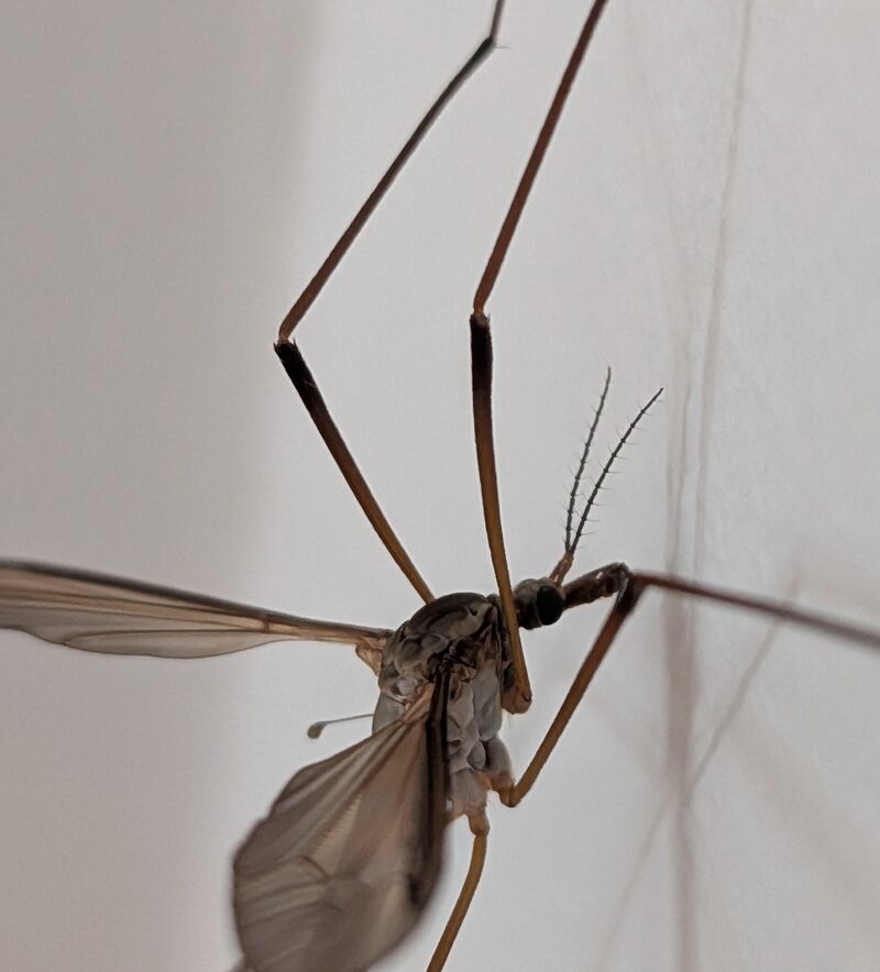
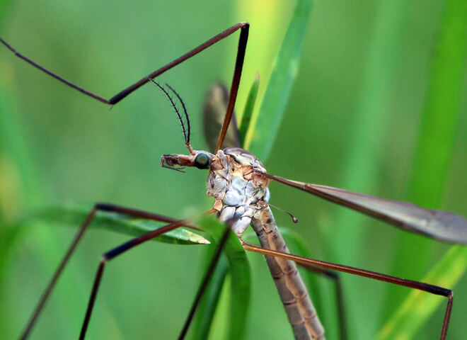
Identified reference photographs of T. paludosa and its closest look-alike,
T. oleracea, against the same views of this specimen. These confirm genus and sex;
they cannot pick the species, and the reason is visible at a glance.
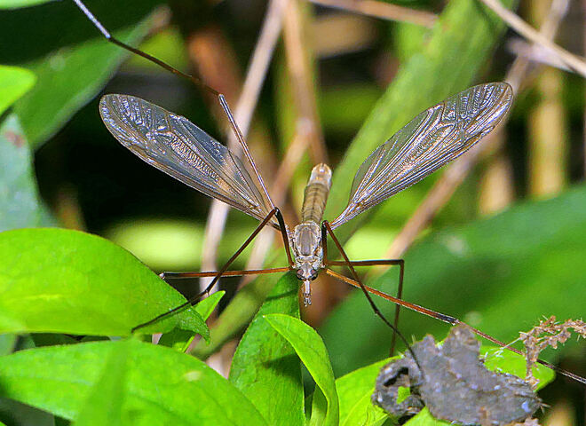
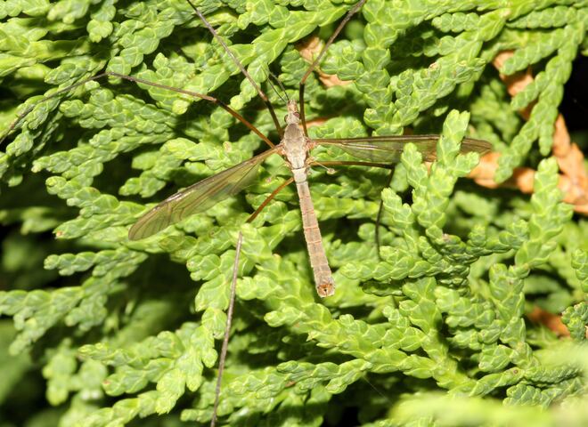
Habitus — the whole animal from above
This specimenCopenhagen, 1 Sept
Grey-brown, plain smoky wings held spread, dark pterostigma, darker stripe along the abdomen.
44 phone photographs of one individual, 09:36–16:03 local time, reviewed as contact sheets before any frame was selected.
Plates are crops upscaled 2–4× with Lanczos resampling; magnification was kept below the point where JPEG artefacts become structure.
Body length estimated by pixel-scaling against a ruler photographed in approximately the same plane.
The antenna was isolated by rotating the whole frame until it lay horizontal, then cropping a band narrow enough to exclude the opposite antenna and its shadow.
Segment boundaries were marked at fixed coordinates and checked against the image rather than counted by eye alone.
Times are local (CEST); the camera names its files in UTC.
No published key was run against the specimen directly; the characters above are drawn
from the secondary sources cited, and the keys listed are what would confirm the
determination.

.jpg)
.jpg)


_head.jpg)